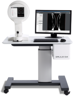
GALILEI G4
|
Reaching a new level in corneal topography and tomography from Ziemer.
The GALILEI G4, is modular and mobile Dual Scheimpflug and Placido system for refractive and cataract surgery. |
Reliable surface data for refractive and corneal surgery
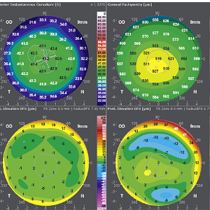
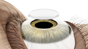
The GALILEI G4 integrates Placido disc topography and Dual Scheimpflug tomography in one device. This combination of technologies allows for a complete analysis of both anterior and posterior corneal surface. The simultaneous recorded Dual Scheimpflug images produce reliable pachymetry and posterior curvature data, whereas the Placido ring images provide highly accurate and central anterior corneal curvature data fitted to the anterior corneal surface. |
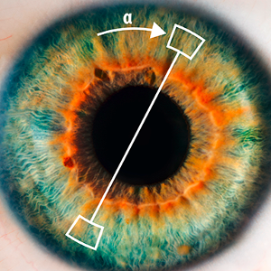
Patented iris-based eye motion compensation
Small to moderate eye motions which can lead to clinically relevant surface curvatures cannot be prevented especially in elderly patients or children. The GALILEI G4 comes with a patented iris tracker which compensates for eye motion. Different than other topography and tomography devices that align data to the pupil or the apex of the cornea, the GALILEI G4 aligns all data to the visual axis using the 1st Purkinje image. This ensures consistent alignment when comparing a series of consecutive measurements over time. |
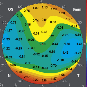
Ray-traced Total Corneal Wavefront
With this powerful ray-traced Total Corneal Wavefront solution, the GALILEI G4 precisely measures high order aberrations for highly predictable outcomes in cataract surgery. The high order aberration display helps identify the most suitable IOL for every patient. |
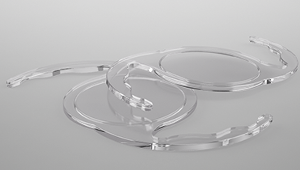
Corneal Implant Planning
The GALILEI G4 comes with a licensable corneal inlay software module which is optimized to patients with implanted corneal inlays. This ensures an accurate and reliable post-op follow-up of those patients. When planning an intracorneal ring surgery, corneal pachymetry, high order aberrations, curvature maps and total corneal astigmatism deliver the information needed to decide on the right ring and corneal position for the treatment. |
Planning and follow-up of Keratoplasty with the GALILEI anterior and posterior corneal astigmatism can be closely controlled. This can be particularly helpful when planning a selective suture removal or in order to follow-up on DSAEK surgery outcomes. High definition corneal pachymetry maps deliver important information about donor tissue cut quality and later visual acuity.
|
Keratoconus Screening
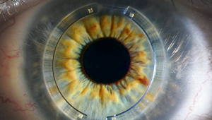
The GALILEI G4 offers a complete dataset for Keratoconus screening. Precise posterior corneal curvature and elevation make it easy to detect posterior corneal bulging and signs of corneal asymmetry even in very early stages. |
IOL Calculation
The GALILEI G4 is a comprehensive tool for IOL planning and calculation. In one measurement, both anterior and posterior corneal data is captured. Data displayed includes anterior and posterior astigmatism, higher order aberrations as well as total corneal power. A licensable IOL calculator integrated the renowned Shammas no-history formula which allows for reliable IOL prediction in post-refractive cases where a patient's clinical history is no longer available. |