System Vision ENG
Info +30 210 4227460
  • Home
  • Company
    • Quality
    • Suppliers
  • Products
    • Imaging Equipment >
      • Slitlamp Imaging
      • Microsurgery Imaging
    • Diagnostic Equipment >
      • BreathPlexi™
      • Auto Ref / Keratometers >
        • VX160
        • VX130
        • VX118
        • VX110
        • ARKM 200
        • L67
        • PLUSOPTIX A12C / A12R
      • Refractive Units >
        • COMBI 400
        • VX 3000
        • VX 2000
        • VX 1100
        • VX 1000
        • ELECTRIC TABLES
      • Refraction Accessories >
        • Trial Frames
        • Optometry Accessories
        • Lenses
      • Chart Displays >
        • VX24
        • VX19
        • CP 40
        • L29i
      • Ultrasounds >
        • AVISO S
        • AVISO
        • AXIS NANO
        • POCKET II
      • Perimeters >
        • PTS 2000
        • PTS 925W
        • PTS-920
      • Slit Lamps >
        • 700GL
        • 30GL
        • 2ZL
        • 4ZL
        • 300XL
      • Tonometers >
        • SNT 700
        • PASCAL
        • AT-1
      • Scheimpflug Topography >
        • VX130
        • GALILEI G6
        • GALILEI G4
      • Placido Topography >
        • VX130
        • VX120
        • KERATRON
        • SCOUT
        • PICCOLO
      • Lensmeters >
        • VX40
        • VX35
        • LM-30
      • Phoropters >
        • VX60
        • VX55
        • VT-5
        • VX50
      • Spectral OCT >
        • REVO NX
        • REVO
    • Lasers >
      • Excimer Lasers >
        • AMARIS 1050RS
        • AMARIS 750S
        • AMARIS 500E
        • SCHWIND CAM
      • Femtosecond Lasers >
        • FEMTO LDV Z8
        • FEMTO LDV Z6 Power Plus
        • FEMTO LDV Z4
        • FEMTO LDV Z2
      • Multispot Lasers >
        • EASYRET Multispot & Subliminal™
        • VITRA 532nm Multispot
      • Monospot Lasers >
        • VITRA 532nm Monospot
      • SLT Lasers >
        • OPTIMIS FUSION
        • SOLUTIS
      • Yag Lasers >
        • OPTIMIS II
        • OPTIMIS FUSION
    • Surgical Equipment >
      • Cross-Linking >
        • CCL VARIO
      • Surgical Microscopes >
        • OM-9 LED
        • OM-5
      • Surgical Furniture >
        • MULTILINE 2
      • Cryosurgery >
        • CRYOLINE
    • IOL's & Implants >
      • Refractive IOL's >
        • ARTISAN PIOL
        • ARTIFLEX PIOL
      • Cataract IOL's >
        • PRECIZON TORIC
        • PRECIZON MONOFOCAL
        • ARTISAN APHAKIA
      • Trauma IOL's >
        • IRIS PROSTHETIC SYSTEM IPS
    • Consumables
  • Special Offers
  • News & Updates
  • Contact
PRODUCTS > DIAGNOSTIC EQUIPMENT > PLACIDO TOPOGRAPHY > VX130

VX130

Picture
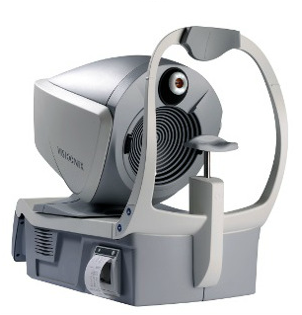
Pre-op and post-op support by a comprehensive COMPLETE ANTERIOR SEGMENT ANALYSIS VX130 from Luneau.

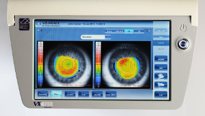
“THE VX130 COMBINES STATE-OF-THE-ART TECHNOLOGIES (SCHEIMPFLUG SCAN CAMERA, NON-CONTACT TONOMETRY, ABERROMETRY, CORNEAL TOPOGRAPHY) AND PROVIDES ESSENTIAL DATA FOR AN IMPROVED TREATMENT OF PATIENTS, REGARDLESS OF THEIR CONDITION AND OCULAR HISTORY. WITH FULLY AUTOMATED MEASUREMENTS, THE VX130 IS THE IDEAL PATIENT MONITORING SYSTEM.”

Complete analysis of the cornea
Combination of data obtained by the Scheimpflug camera and corneal topography data, thickness maps and elevation maps can be obtained on a broad corneal surface.

  • Corneal thickness  map
  • Elevation  map
  • Anterior and posterior axial, tangential and refraction maps
  • Anterior and posterior keratometry, eccentricity

PRE-OP cataract surgery
  • Visualization of crystalline  opacities
  • Analysis of wavefront aberrations, with the ability to separate corneal and lenticular/internal aberrations

POST-OP cataract surgery
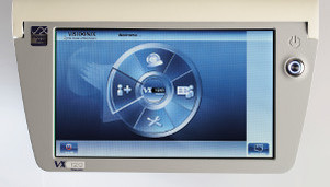
  • Post-op check on intraocular lens implants
  • Axis alignment  check of the toric lens implant
  • Analysis of post-op output to improve surgery protocol

Picture
Touch screen
VX130 has a large integrated color touch screen 10.1" with recliner for handling the device by sitting or standing.
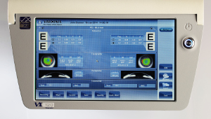
Picture
Measurements
  •      Refractometry
  •      Keratometry
  •      Aberrometry
  •      Corneal Topography
  •      Accommodation
  •      Pupilometry
  •      Tonometry
  •      Pachymetry & Anterior Chamber (with    Scheimpflug technology)
Picture
Objective refraction and analysis of aberrations
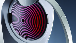
  • 1200 points of analysis for a pupil of 7 mm in diameter
  • Objective refraction under mesopic and photopic conditions
  • Measures lower-order and higher-order aberrations
  • Access visual acuity and quality of vision from a pupil diameter as small as 1.2 mm
Picture
Corneal Topography
  • Axial, Tangential, Elevation and refraction maps
  • Keratometry
  • Contact lens fitting
  • Keratoconus  screening
  • Eccentricity
  • Corneal aberrometry


Picture
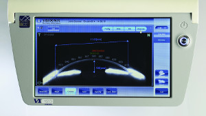
Tonometry / Pachymetry / Irido-Corneal Angles
  • Measurement  of IOP (intraocular pressure)
  • Measurement  of corneal thickness using Scheimpflug imaging
  • Corrected  IOP as a function of corneal thickness
  • Automatic measurement  of irido-corneal angles using Scheimpflug imaging


Picture
Tonometry
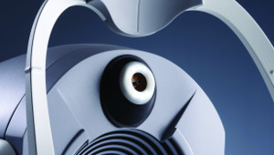
Fully automated non-contact tonometry procedure. A soft
air puff is measured the pressure preventing discomfort to the patient. Accuracy in measurement and display of the true IOP according with the pachymetry.
Picture
Printing
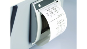
The device has a built-in thermal printer for printing the results of measurement for refractometric and keratometry. For the printing of topographic maps it is possible by connecting an external printer.
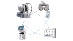
Picture
VX Network
The VX130 easily connects through wireless or wired  diagnostic devices of Visionix. The phoropter (VX60, VX55), lensmeter (VX40, VX35) and the chart monitor (VX24, VX19) create a network with the help of VX BOX which transfers the data between the devices and also sends the data to a EMR software.
Copyright © 2000-2019 System Vision SA | All rights reserved
Greek
English
Terms of Use