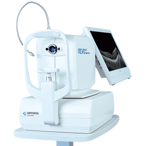
REVO OCT Copernicus
|
Our supreme experience in Spectral Domain OCT allows us to provide the market updated SOCT Copernicuse REVO with next generation spectrometer offering noticeable faster scan speed and enhance image quality through the whole scanning window.
SOCT Copernicus REVO meets all demands in daily routine practice. The main feature of updated REVO is increased scanning speed to 80 000 A-scan/sec. The updated SOCT Copernicus REVO is available with the Angiography module. Higher scanning speed reduce scan time to minimize motion artefacts. Higher sensitivity spectrometer allows to visualize finer details. Key improvements are:
|
OCT made simple as never before. All it must be done is to position the patient and press the START button to acquire examinations of both eyes. The device will make examination independently.
Small system footprint, various operator and patient positions allow to install SOCT Copernicus REVO even in the smallest examination room. Variety of review and analysis tool give the operator a choice of using it as a screening or as an advanced diagnostic device.
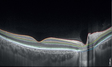
The noise reduction technology provides the finest details proven to be important for early disease detection.
Small system footprint, various operator and patient positions allow to install SOCT Copernicus REVO even in the smallest examination room. Variety of review and analysis tool give the operator a choice of using it as a screening or as an advanced diagnostic device.
The noise reduction technology provides the finest details proven to be important for early disease detection.

|
|
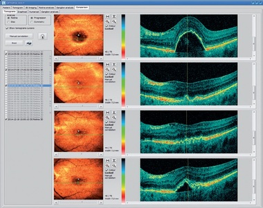
Single 3D Retina examination is enough to perform both Retina and Glaucoma analysis based on retinal scans.
Software automatically recognizes 8 retina layers, thus allowing a more precise diagnosis and mapping of any changes in the patient’s retina condition.
Software automatically recognizes 8 retina layers, thus allowing a more precise diagnosis and mapping of any changes in the patient’s retina condition.
|
|
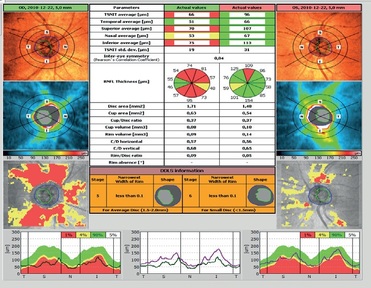
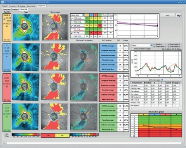
Comprehensive glaucoma analysis tools for Quantification of Optic Nerve Head, Retina Nerve Fiber Layer, DDLS, Ganglion layer and Asymmetry
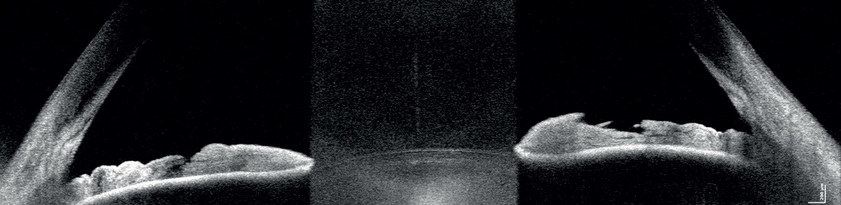
For standard examinations no additional lens is required. Additional adapter provided with the device allows to make wide scans of anterior segment.
|
|
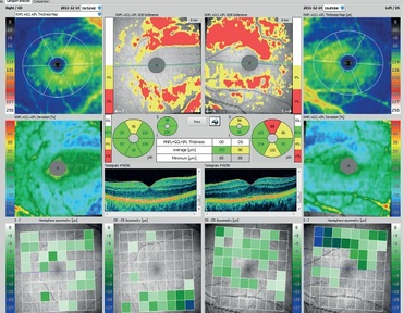
High density of standard 3D scan allows to precisely track the disease progress. Operator can analyze changes in morphology, quantified progression maps or evaluate the progression trends.
A proficient networking solution increases productivity and an enhanced patient experience. It allows you to view and manipulate multiple examinations from review stations in your practice. Effortlessly helping to facilitate patient education by allowing you to interactively show examination results to patients. Every practice will have different requirements which we can provide by tailoring a bespoke service. There is no additional charge for the server module.