REVO NX
|
REVO NX engineering team, designers of the first commercially available Spectral Domain OCT in the World, is proud to present the World’s fastest OCT from Optopol.
Our supreme experience in Spectral Domain OCT allows us to provide the market with the state of the art instrument, offering advanced technologies and remarkable simplicity of operation. The new REVO NX software meets all demands of a daily routine in a modern ophthalmic practice. The new angiography module expands the precision of your diagnosis with a minimum patients fatigue. |
Need for speed
The world’s fastest available scanning speed allows for more achievable and more detailed exams with reduction of the scanning time. It brings benefits for both clinicians and patients by reducing errors often caused by involuntary eye movements.
OCT made simple as never before
Position the patient and press the START button to acquire examinations of both eyes. The REVO NX, using vocal messages, guides the patient through the process, increasing comfort and reducing patient chair time. Short scanning time ensures less fatigue for the patient. Creating customized scanning protocols of different diagnostic scenarios speed’s up the workflow.
A perfect fit for every practice.
With a small system footprint and access for both operator and patient from one side, space saving is further enhanced. In addition, connection by a single cable allows the installation of REVO NX into the smallest of examination rooms. REVO NX's variety of examination and analysis tools enables it to effortlessly function as a screening or advanced diagnostic device.
High quality of OCT image
The noise reduction technology provides the finest details proven to be important for early disease detection.
|
|
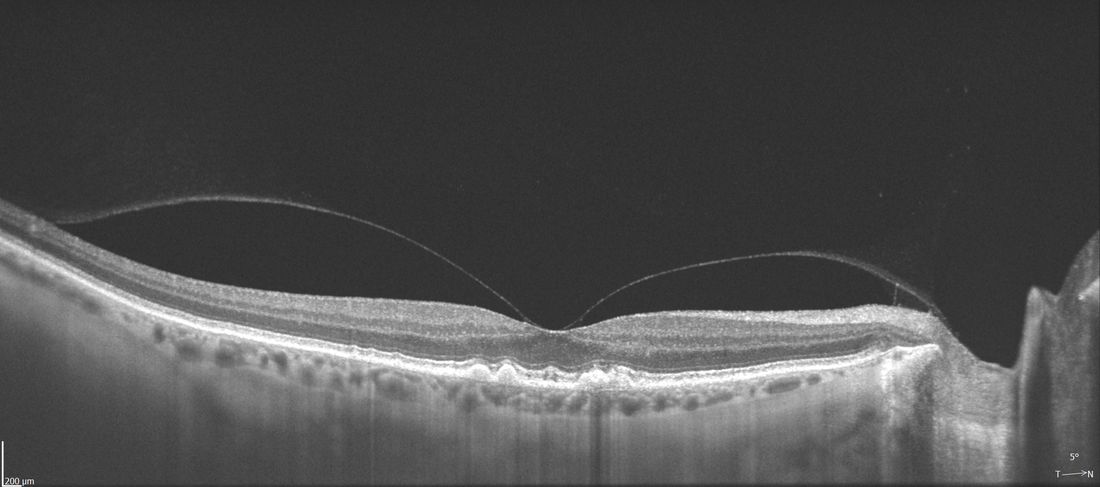
RETINA
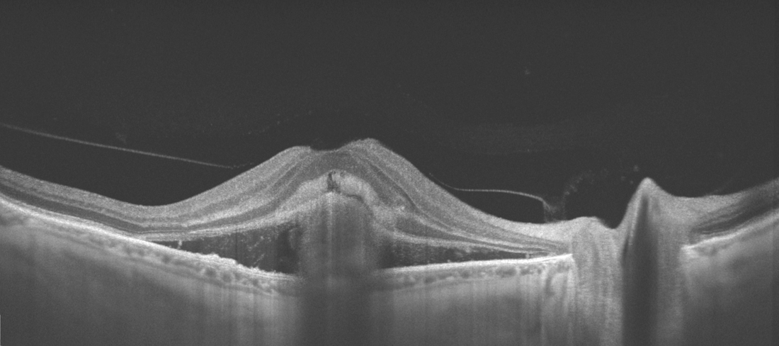
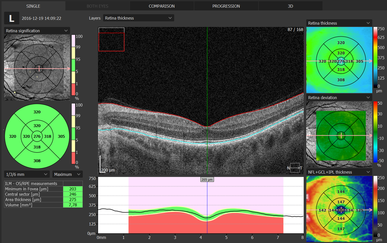
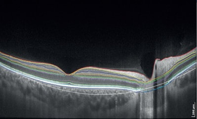
Single 3D Retina examination is enough to perform both Retina and Glaucoma analysis based on retinal scans. Software automatically recognize 8 retina layers. Thus allowing a more precise diagnosis and mapping of any changes in the patient’s retina condition.
Wide field Scan
12x12 mm wide field Central scan is perfect for fast and precise screening of the patient’s retina. Dense scanning in high resolution tomograms guarantee most of the discoveries of early changes. Peripheral scanning reveals diseases in the far periphery.
|
|
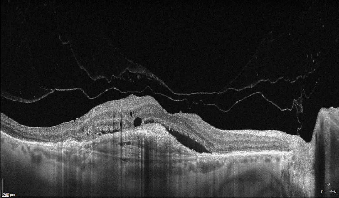
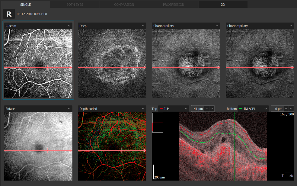
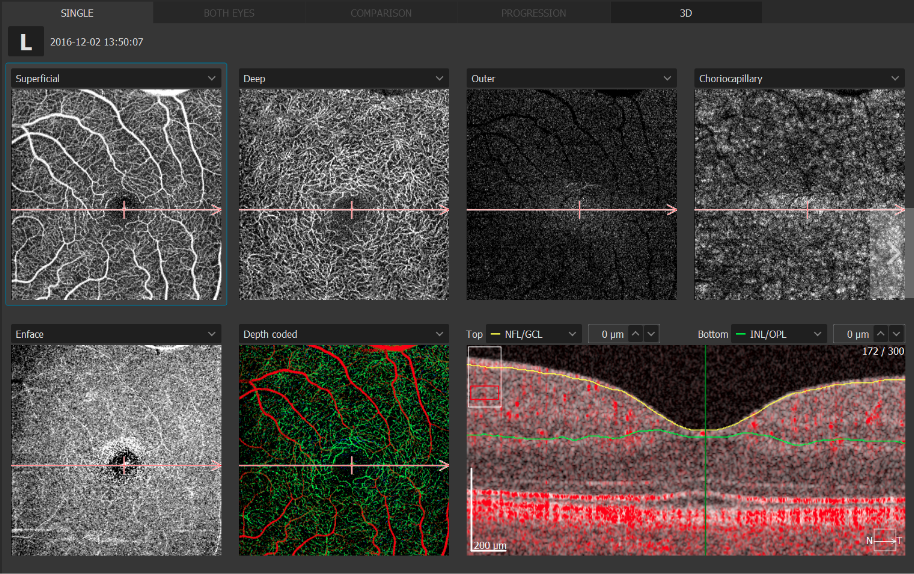
Angiography OCT
This non-invasive dye free technique allows the visualization of the microvasculature of the retina. Both blood flow and structural visualization will give additional information in the diagnosis of many retinal diseases. Angiography scan allows assessment of the structural vasculature of the macula, periphery or the optic disc. Extremely short scanning time 1.6 second in standard resolution or in high resolution within ~3 seconds.
This non-invasive dye free technique allows the visualization of the microvasculature of the retina. Both blood flow and structural visualization will give additional information in the diagnosis of many retinal diseases. Angiography scan allows assessment of the structural vasculature of the macula, periphery or the optic disc. Extremely short scanning time 1.6 second in standard resolution or in high resolution within ~3 seconds.
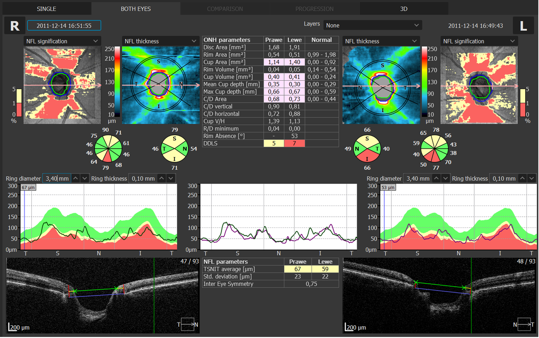
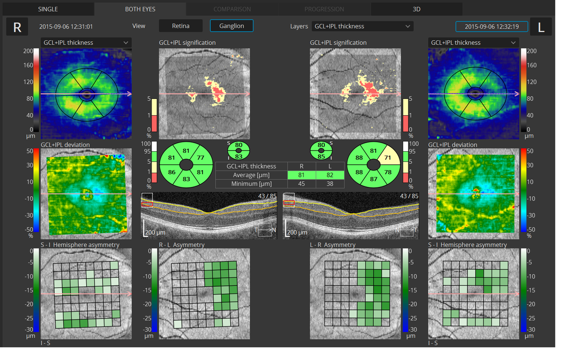
Glaucoma
Comprehensive glaucoma analysis tools for quantification of the Nerve Fiber Layer, Ganglion layer and Optic Head with DDLS allows for precise diagnosis and the monitoring of glaucoma over time. Asymmetry Analysis of Ganglion layers between hemispheres and between eyes allows easier identification and detection of glaucoma in early stages and in non-typical patients.
Comprehensive glaucoma analysis tools for quantification of the Nerve Fiber Layer, Ganglion layer and Optic Head with DDLS allows for precise diagnosis and the monitoring of glaucoma over time. Asymmetry Analysis of Ganglion layers between hemispheres and between eyes allows easier identification and detection of glaucoma in early stages and in non-typical patients.
|
|
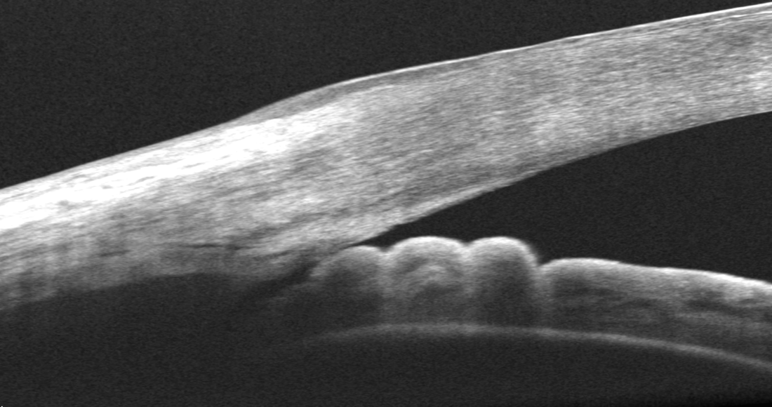
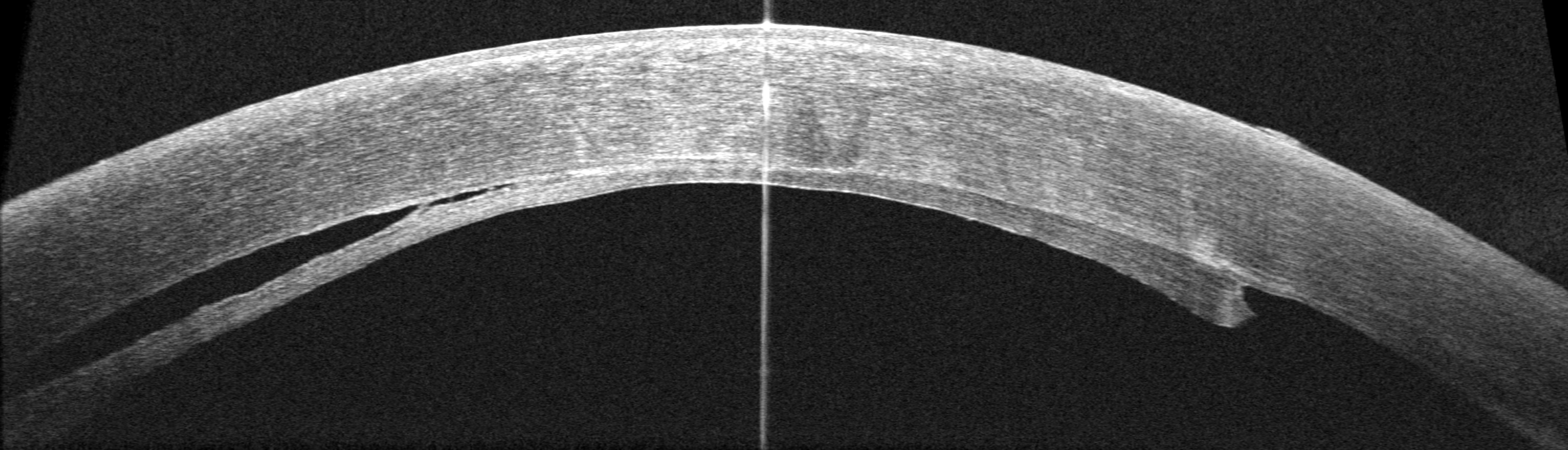
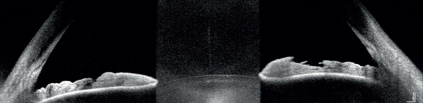
ANTERIOR
For a standard anterior examination, no additional lens is required. This allows the examiner to quickly complete the scanning procedure. Presentation of results for both eyes allows quick and precise evaluation of the condition of the anterior segment.
Additional adapter provided with the device increases range of clinical application in Anterior chamber observation
FOLLOW UP
REVO NX's standard high density scanning capability and blood vessel structure recognition enables a precise alignment of past and current scans. The Operator can analyze changes in morphology, quantified progression maps and evaluate the progression trends.
NETWORKING
A proficient networking solution increases productivity and an enhanced patient experience. It allows you to view and manipulate multiple examinations from review stations in your practice. Effortlessly helping to facilitate patient education by allowing you to interactively show examination results to patients. Every practice will have different requirements which we can provide by tailoring a bespoke service. There is no additional charge for the server module.